Antioxidant evaluation and bio-guided isolation from methanol leaf extract of Acalypha godseffiana

Authors

  • S. D. Umoh Department of Chemistry, Department of Chemistry, Joseph Sarwuan Tarka University, Makurdi, Nigeria
  • A. K. Asekunowo Department of Chemistry, Faculty of Science, University of Lagos, Nigeria
  • I. S. Okoro Department of Chemistry, Department of Chemistry, Joseph Sarwuan Tarka University, Makurdi, Nigeria
  • N. X. Siwe Department of Chemistry, Faculty of Science, Rhodes University, South Africa
  • R. W. M. Kraus Department of Chemistry, Faculty of Science, Rhodes University, South Africa
  • O. O. Okoh Department of Chemistry, Faculty of Science and Agriculture, University of Fort Hare, Alice South Africa
  • A. O. T. Ashafa Department of Plant Science, Faculty of Natural and Agricultural Science, University of the Free State, South Africa
  • O. T. Asekun Department of Chemistry, Faculty of Science, University of Lagos, Nigeria
  • O. B. Familoni Department of Chemistry, Faculty of Science, University of Lagos, Nigeria

Keywords:

Acalypha godseffiana, spectroscopy, antioxidant, antifungal, bioactive compounds

Abstract

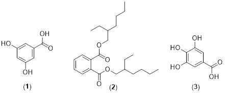
Acalypha godesffiana is a plant used in conventional medicine for fungal-related illnesses. The plant‘s extracts were investigated in this study, antioxidant, and antimicrobial studies were conducted. Different models were employed in the antioxidant assay; serial dilution was utilized to determine the minimum inhibitory concentration (MIC). The extract from A. godseffiana was purified and characterized by chromatographic and spectroscopic techniques, respectively. Three biologically active compounds, 3, 5-dihdroxylbenzoic acid (1), 3, 4, 5-trihydroxybenzoic acid (2), as well as Di-(2-ethylhexyl) phthalate (3), were identified for the first time in A. godseffiana. The extract and fractions exhibited varying scavenging capacities on different anti-oxidative models. The DPPH of MeOH (IC50= 0.51 mg mL^-1) was comparable with silymarine (SLY) IC50= 0.52 mg mL^-1 and better than gallic acid (GAL) IC50=1.95 mg mL^-1; the ABTS.+ of EtOAc column fraction (ACF, IC50=0.46 mg mL^-1) was comparable with standard SLY, IC50 = 0.47 mg mL^-1; and an OH radical of DCM, IC50= 0.10 mg mL^-1, was better than both standards (SLY, IC50= 6.30 mg mL^-1, GAL: IC50= 1.93 mg mL^-1). ACF showed superior antifungal activities (0.02 mg mL^-1) against Cryptococcus neoformans and Candida albicans, compared to ketoconazole (MIC of 0.250 mg mL^-1). Compounds (1-3) from A. godseffina reportedly displayed antioxidant and other activities. This study validated the antifungal potentials of A. godseffiana leaves and identified bioactive compounds. The extracts should be further investigated, and the compounds should be added to the existing library for further investigation of possible leads.


Dimensions

M. Ahvazi, F. Khalighi-Sigaroodi, M. M. Charkhchiyan, F. Mojab, V. A. Mozaffarian & H. Zakeri, “Introduction of medicinal plants species with the most traditional usage in Alamut region”, Iranian Journal of Pharmaceutical Research 11 (2012) 185. https://www.ncbi.nlm.nih.gov/pmc/articles/PMC3813099/.

G. M. Cragg, P. G. Grothaus & D. J. Newman, “New horizons for old drugs and drug leads”, Journal of Natural Products 779 (2014) 703. https://pubs.acs.org/doi/abs/10.1021/np5000796.

M. S. Butler, “The role of natural product chemistry in drug discovery”, Journal of Natural Products 67 (2004) 2141. https://pubs.acs.org/doi/abs/10.1021/np040106y.

D. Lawal & I. Yunusa, “Dodonea Viscosa Linn: its medicinal, pharmacological and phytochemical properties”, International Journal of Innovation and Applied Studies 2 (2013) 476.

https://www.researchgate.net/publication/281956749_Dodonea_Viscosa_Linn_Its_Medicinal_Pharmacological_and_Phytochemical_Properties.

K. Lewis & F. M. Ausubel, “Prospects for plant-derived antibacterials”, Nature Biotechnology 24 (2006) 1504. https://www.nature.com/articles/nbt1206-1504.

A. Pandey & S. K. Shashank Kumar, “Perspective on plant products as antimicrobials agents: a review”, Pharmacologia 4 (2013) 469. https://www.cabidigitallibrary.org/doi/full/10.5555/20143165833.

J. H. Doughari, Phytochemicals: extraction methods, basic structures and mode of action as potential chemotherapeutic agents, INTECH Open Access Publisher Rijeka, Croatia,2012, pp. 1–33. https://chemical.report/Resources/Whitepapers/022971ea-d752-4f91-b6dd-9c0f567501ec whitepaper1.pdf.

J. Parekh & S. Chanda, “Antibacterial and phytochemical studies on twelve species of Indian medicinal plants”, African Journal of Biomedical Research 10 (2007) 175. https://tspace.library.utoronto.ca/handle/1807/54086.

O. Kingsley, A. A. Marshall & I. I. Inegbenose, “Phytochemical, proximate and elemental analysis of Acalypha wilkesiana leaves”, Scientific Journal of Pure and Applied Sciences 2 (2013) 323.

https://www.researchgate.net/publication/307695658_Phytochemical_proximate_and_elemental_analysis_of_acalypha_wilkesiana_eaves.

A. Asekunowo, A. Ashafa, O. Okoh, O. Asekun & O. Familoni, “Evaluation of Phytochemical Constituents and Antifungal Properties of Different Solvent Extracts of the Leaf of Acalypha godseffiana Mull Arg”, University of Lagos Journal of Basic Medical Sciences 5(0) (2021).

https://jbms.unilag.ng/index.php/jbms/article/viewFile/20/20.

R. Jayaprakasam & T. Ravi, “Evaluation of anti arthritic activity of the root extract of Acalypha indica Linn. using in vitro techniques”, Int J Phyto Pharm 2 (2012) 169. https://journals.indexcopernicus.com/search/article?articleId=606664.

V. Takle, R. Savad, A. Kandalkar, A. Akarte & A. Patel, “Pharmacognos tic and Phytochemical investigations of aerial parts of Acalypha indica

Linn”, Pharmacognosy Journal 3 (2011) 33. https://doi.org/10.5530/pj.

21.6.

M. A. El-raey, T. K. Mohamed, W. A. El-kashak & W. O. Fayad, “Phenolic constituents and biological activities of Acalypha wilkesiana forma tricolor muell arg seeds”, International Journal of Pharmacognosy and Phytochemical Research 8 (2016) 386. https://www.researchgate.net/profile/

Mohamed-El-Raey-2/publication/299424406.

O. H. Oladimeji, E. U. Tom & E. E. Attih, “Ethyl gallate and pyrogallol from Acalypha wilkesiana var. Lace-acalypha (Muell & Arg.)”, European Chemical Bulletin 3 (2014) 788. https://www.cabidigitallibrary.org/doi/full/10.5555/20143380277.

N. Okarter, C. S. Liu, M. E. Sorrells & R. H. Liu, “Phytochemical content and antioxidant activity of six diverse varieties of whole wheat”, Food Chemistry 119 (2010) 249. https://doi.org/10.1016/j.foodchem.2009.06.021.

S. Silici, O. Sagdic & L. Ekici, “Total phenolic content, antiradical, antioxidant and antimicrobial activities of Rhododendron honeys”, Food chemistry 121 (2010) 238. https://doi.org/10.1016/j.foodchem.2009.11.078.

A. Floegel, D. O. Kim, S. J. Chung, S. I. Koo & O. K. Chun, “Comparison of ABTS/DPPH assays to measure antioxidant capacity in popular antioxidant-rich US foods”, Journal of Food Composition and Analysis

(2011) 1043. https://doi.org/10.1016/j.jfca.2011.01.008.

L. Muller, K. Fr ¨ ohlich & V. B ¨ ohm, “Comparative antioxidant activities of carotenoids measured by ferric reducing antioxidant power (FRAP), ABTS bleaching assay (αTEAC), DPPH assay and peroxyl radical scavenging assay”, Food Chemistry 129 (2011) 139. https://doi.org/10.1016/j.foodchem.2011.04.045.

H. Ramakrishna, S. S. Murthy, R. Divya, D. MamathaRani & M. G.Panduranga, “Hydroxy radical and DPPH scavenging activity of crude protein extract of Leucas linifolia: A folk medicinal plant”, Asian Journal

of Plant Science and Research 2 (2012) 30. https://www.academia.edu/4946331/Hydroxy radical and DPPH scavenging activity of crude protein extract of Leucas linifolia A folk medicinal plant.

M. O. Sofidiya & O. B. Familoni, “In vitro antioxidant activities of ethanolic stem bark extract of Brachystegia eurycoma”, University of Lagos Journal of Basic Medical Sciences 4 (2016) 31. https://ir.unilag.edu.ng/items/b1a8e1a8-466d-44f3-b276-aaa6e3acaaeb.

A. O. T. Ashafa, “Medicinal potential of Morella serata (Lam.) Killick (Myricaceae) root extracts: biological and pharmacological activities”, BMC Complementary and Alternative Medicine 13 (2013) 1. https://link.springer.com/article/10.1186/1472-6882-13-163.

A. A. Mohdaly, I. Smetanska, M. F. Ramadan, M. A. Sarhan & A. Mah moud, “Antioxidant potential of sesame (Sesamum indicum) cake extract in stabilization of sunflower and soybean oils”, Industrial Crops and Products 34 (2011) 952. https://doi.org/10.1016/j.indcrop.2011.02.018.[23] B. L. Halvorsen, M. H. Carlsen, K. M. Phillips, S. K. Bøhn, K. Holte, D. R. Jacobs Jr & R. Blomhoff, “Content of redox-active compounds (ie, antioxidants) in foods consumed in the United States”, The American journal of clinical nutrition 84 (2006) 95. https://doi.org/10.1093/ajcn/84.1.95.

K. Sowndhararajan & S. C. Kang, “Free radical scavenging activity from different extracts of leaves of Bauhinia vahlii Wight & Arn”, Saudi Journal of Biological Sciences 20 (2013) 319. https://doi.org/10.1016/j.sjbs.2012.12.005.

P. Prieto, M. Pineda & M. Aguilar, “Spectrophotometric quantitation of antioxidant capacity through the formation of a phosphomolybdenum complex: specific application to the determination of vitamin E”, Analytical Biochemistry 269 (1999) 337. https://doi.org/10.1006/abio.1999.4019.

M. O. Sofidiya & O. Familoni, “Antioxidant activities of different solvent extracts of leaves and root of Flabellaria paniculata Cav.(Malpighiaceae)”, Journal of Medicinal Plants Research 6 (2012) 4682. https://ir.unilag.edu.ng/items/3bb664aa-695d-4b4b-aa1c-78463d90429b.

W. Jiang, Y. Qiu, Y. Ni, M. Su, W. Jia & X. Du, “An automated data analysis pipeline for GC− TOF− MS metabonomics studies”, Journal of Proteome Research 9 (2010) 5974. https://doi.org/10.1021/pr1007703.

G. Corea, E. Fattorusso, V. Lanzotti, R. Capasso & A. A. Izzo, “Antispasmodic saponins from bulbs of red onion, Allium cepa L. var. Tropea”, Journal of Agricultural and Food Chemistry 53 (2005) 935. https://pubs.acs.org/doi/abs/10.1021/jf048404o.

F. A. Al-Bayati & H. F. Al-Mola, “Antibacterial and antifungal activities of different parts of Tribulus terrestris L. growing in Iraq”, Journal of Zhejiang University Science B 9 (2008) 154. https://link.springer.com/article/10.1631/jzus.B0720251.

S. D. Umoh, G. Bojase, I. B. Masesane, D. Loeto & R. T. Majinda, “A comprehensive review of Combretum flavonoids and their biological activities: An update between 1990 and 2022”, Biochemical Systematics and Ecology 108 (2023) 104644. https://doi.org/10.1016/j.bse.2023.

S. Kumar & A. K. Pandey, “Chemistry and biological activities of flavonoids: an overview”, The Scientific World Journal 2013 (2013) 16. https://www.hindawi.com/journals/tswj/2013/162750/.

Z. Sroka & W. Cisowski, “Hydrogen peroxide scavenging, antioxidant and anti-radical activity of some phenolic acids”, Food and Chemical Toxicology 41 (2003) 753. https://doi.org/10.1016/S0278-6915(02)00329-0.

J. Nair, A. Ndhlala, J. Chukwujekwu & J. Van Staden, “Isolation of di (2-ethylhexyl) phthalate from a commercial South African cognate herbal mixture”, South African Journal of Botany 80 (2012) 21. https://doi.org/10.1016/j.sajb.2012.01.008.

K. H. Lee, J. H. Kim, D. S. Lim & C. H. Kim, “Anti-leukaemic and antimutagenic effects of di (2-ethylhexyl) phthalate isolated from Aloe vera Linne”, Journal of Pharmacy and Pharmacology 52 (2000) 593. https://doi.org/10.1211/0022357001774246.

M. R. Habib & M. R. Karim, “Antimicrobial and cytotoxic activity of di-(2-ethylhexyl) phthalate and anhydrosophoradiol-3-acetate isolated from Calotropis gigantea (Linn.) flower”, Mycobiology 37 (2009) 31. https://www.tandfonline.com/doi/abs/10.4489/MYCO.2009.37.1.031.

A. Abri & M. Maleki, “Isolation and identification of gallic acid from the elaeagnus angustifolia leaves and determination of total phenolic, flavonoids contents and investigation of antioxidant activity”, Iranian

Chemical Communication 4 (2016) 146. https://icc.journals.pnu.ac.ir/article 1892 8396d285635b52f6ba3bd74ef1e0fc09.pdf.

N. S. Ghaly, M. Nabil, M. H. Grace & F. R. Melek, “Pendulaosides A and B. Two acylated triterpenoid saponins from Harpullia pendula seed extract”, Phytochemistry Letters 21 (2017) 278. https://doi.org/10.1016/j.phytol.2017.06.016.

H. M. Chen, Y. C. Wu, Y. C. Chia, F. R. Chang, H. K. Hsu, Y. C. Hsieh, C. C. Chen & S. S. Yuan, “Gallic acid, a major component of Toona sinensis leaf extracts, contains a ROS-mediated anti-cancer activity in human prostate cancer cells”, Cancer Letters 286 (2009) 161.

https://doi.org/10.1016/j.canlet.2009.05.040.

Compounds identified from Acalypha godseffiana leaf extract.

Published

2024-06-19

How to Cite

Antioxidant evaluation and bio-guided isolation from methanol leaf extract of Acalypha godseffiana. (2024). Journal of the Nigerian Society of Physical Sciences, 6(3), 2038. https://doi.org/10.46481/jnsps.2024.2038

How to Cite

Antioxidant evaluation and bio-guided isolation from methanol leaf extract of Acalypha godseffiana. (2024). Journal of the Nigerian Society of Physical Sciences, 6(3), 2038. https://doi.org/10.46481/jnsps.2024.2038