Synthesis and in vitro bioactivity of sodium metasilicate-derived silicon-substituted hydroxyapatite

Authors

  • Enobong R. Essien Department of Chemical Sciences, Bells University of Technology, Ota 112103, Nigeria https://orcid.org/0000-0003-2379-3640
  • Violette N. Atasie Department of Chemical Sciences, Bells University of Technology, Ota 112103, Nigeria
  • Ngozi A. Adeleye Department of Chemical Sciences, Bells University of Technology, Ota 112103, Nigeria
  • Luqman A. Adams Deparment of Chemistry, University of Lagos, Akoka, Yaba100213, Nigeria

Keywords:

Silicon-substituted hydroxyapatite, Bioactivity, Sodium metasilicate, Bone repair, Synthetic bone grafts

Abstract

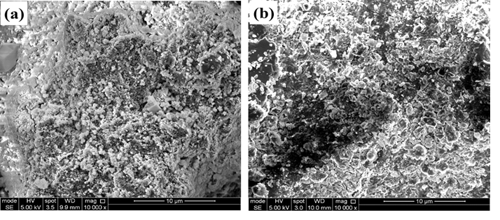
Structural alteration of synthetic implants aims to achieve better bioactivity, higher cellular response, and regulated degradability, all of which are critical criteria for a biomaterial to serve as a graft in bone regeneration. The aim of this work was to synthesize silicon-substituted hydroxyapatite and test its bioactivity in simulated body fluid (SBF) by proving the use of sodium metasilicate (Na2SiO3.9H2O) as an affordable precursor of silica. Thus, the study evaluated the in vitro bone-bonding capacity of hydroxyapatite (Ca10(PO4)6(OH)2) (HA) substituted with silicate ion (Ca10(PO4)6-x(SiO4)x(OH)2-x; SixHA). The SixHA with x = 0.4 was synthesized by utilizing a wet precipitation method with sodium metasilicate as a low-cost silica alternative for alkoxysilane precursors. The SixHA was then examined for properties such as morphology, elemental composition, phase composition, and the nature of chemical bonds using scanning electron microscopy (SEM), energy dispersive X-ray analysis (EDX), X-ray diffractometry (XRD), and Fourier transformed infrared spectroscopy (FTIR), respectively. An in vitro bioactivity experiment was also carried out by incubating the SixHA in simulated body fluid (SBF) at 36.5 °C for 7 and 14 days. The obtained results revealed the substitution of SiO44- for some PO43- groups in the hydroxyapatite structure. The SixHA nucleated more apatite crystals on its surface and demonstrated some degradability during the periods of immersion in SBF. The characteristics of the SixHA imply that it could be used as a graft in bone restoration applications, thus signifying that sodium metasilicate could serve as an economic silica source for silicon-substituted hydroxyapatite production. 

Dimensions

S. Daane, “Alloplastic implantation”, in Plastic Surgery Secrets Plus, J. Weinzweig (Ed.), Mosby, St. Louis, United States, 2010, pp. 28–32. https://doi.org/10.1016/B978-0-323-03470-8.00005-3.

H. A. Siddiqui, K. L. Pickering, & M. R. Mucalo, “A Review on the use of hydroxyapatite-carbonaceous structure composites in bone replacement materials for strengthening purposes”, Materials (Basel) 11 (2018) 1813. https://doi.org/10.3390/ma11101813.

A. Camaioni, I. Cacciotti, L. Campagnolo, & A. Bianco, “Siliconsubstituted hydroxyapatite for biomedical applications”, in Hydroxyapatite (HAp) for Biomedical Applications, M. Mucalo (Ed.), Woodhead Publishing, Cambridge, England, 2015, pp. 343–373. https://doi.org/10.1016/B978-1-78242-033-0.00015-8.

J. H. Shepherd, D.V. Shepherd, & S. M. Best, “Substituted hydroxyapatites for bone repair”, Journal of Materials Science: Materials in Medicine 23 (2012) 2335. https://doi.org/10.1007/s10856-012-4598-2.

M. Vallet-Reg´?, & J. M. Gonzalez-Calbet, “Progress in Solid State Chem-´ istry”, 32 (2004) 1. https://doi.org/10.1016/j.progsolidstchem.2004.07.001.

R. Z. LeGeros, “Properties of osteoconductive biomaterials: Calcium phosphates”, Clinical Orthopaedics and Related Research 395 (2002) 81. https://doi.org/10.1097/00003086-200202000-00009.

S. V. Dorozhkin, & M. Epple, “Biological and medical significance of calcium phosphates”, Angewandte Chemie International Edition 41 (2002) 3130. https://doi.org/10.1002/1521-3773(20020902)41:17?3130::AID-ANIE3130?3.0.CO;2-1

S. V. Dorozhkin, “Bioceramics based on calcium orthophosphates. (Review)”, Glass Ceramics 64 (2007) 442. https://doi.org/10.1007/s10717-007-0109-1.

E. M. Carlisle, “Silicon: A possible factor in bone calcification”, Science 167 (1970) 279. https://doi.org/10.1126/science.167.3916.279.

E. M. Carlisle, “Silicon: an essential element for the chick”, Science 178 (1972) 619. https://doi.org/10.1126/science.178.4061.619.

E. M. Carlisle, “Silicon: a requirement in bone formation independent of vitamin D1”, Calcified Tissue International 33 (1981) 27. https://doi.org/10.1007/BF02409409.

E. M. Carlisle, “Silicon as an essential trace element in animal nutrition”, Ciba Foundation Symposium 121 (1986) 123. https://doi.org/10.1002/9780470513323.ch8.

K. Schwarz, “A bound form of silicon in glycosaminoglycans and polyuronides”, Proceedings of the National Academy of Sciences, USA 70 (1973) 1608. https://doi.org/10.1073/pnas.70.5.1608.

M. Calomme, P. Geusens, N. Demeester, G. J. Behets, P. D’Haese, J. B. Sindambiwe, V. Van Hoof, & D. Vanden Berghe, “Partial prevention of long-term femoral bone loss in aged ovariectomized rats supplemented with choline-stabilized orthosilicic acid”, Calcified Tissue International 78 (2006) 227. https://doi.org/10.1007/s00223-005-0288-0.

B. D. Nielsen, G. D. Potter, E. L. Morris, T. W. Odom, D. M. Senor, J. A. Reynolds, W. B. Smith, M. T. Martin, & E. H. Bird, “Training distance to failure in young racing quarter horses fed sodium zeolite”, Journal of Equine Veterinary Science 13 (1993) 562. https://doi.org/10.1016/S0737-08060681526-1.

I. R. Gibson, S. M. Best, & W. Bonfield, “Chemical characterization of silicon-substituted hydroxyapatite”, Journal of Biomedical Materials Research 44 (1999) 422. https://doi.org/10.1002/(sici)1097-4636(19990315)44:4?422::aid-jbm8?3.0.co;2-#

A. J. Ruys “Silicon-doped hydroxyapatite”, Journal of the Australian Ceramic Society 29 (1993) 71. https://www.docketalarm.com/cases/PTAB/IPR2013-00582/Inter_Partes_Review_of_U.S._Reissue_Pat._RE041251/09-12-2013-Petitioner/Exhibit-1014-AJ_Ruys,_Silicon_Doped_Hydroxyapatite/.

A. A. Faremi, S. S. Oluyamo, K. D. Adedayo, Y. A. Odusote, & O. I. Olusola, “Influence of silicon nanoparticle on the electrical properties of heterostructured CdTe/CdS thin films based photovoltaic device”, Journal of the Nigerian Society of Physical Sciences 3 (2021) 256. https://doi.org/10.46481/jnsps.2021.267.

K. M. Omatola, A. D. Onojah, A. N. Amah, & I. Ahemen, “Synthesis and characterization of silica xerogel and aerogel fromrice husk ash and pulverized beach sand via sol-gel route”, Journal of the Nigerian Society of Physical Sciences 5 (2023) 1609. https://doi.org/10.46481/jnsps.2023.5.1609.

D. Arcos, J. Rodriguez-Carvajal, & M. Vallet-Regi, “The effect of the silicon incorporation on the hydroxylapatite structure. a neutron diffraction study”, State Sciences 6 (2004) 987. https://doi.org/10.1016/j.solidstatesciences.2004.05.001.

Y. Tanizawa, & T. Suzuki, “X-ray photoelectron spectroscopy study of silicate-containing apatite, Phosphorus”, Phosphorus Research Bulletin 4 (1994) 83. https://doi.org/10.3363/prb1992.4.083.

X.L. Tang, X.F. Xiao, & R.F. Liu, “Structural characterization of siliconsubstituted hydroxyapatite synthesized by a hydrothermal method”, Materials Letters 59 (2005) 3841. https://doi.org/10.1016/j.matlet.2005.06.060.

D. Arcos, J. Rodriguez-Carvajal, & M. Vallet-Regi, “Silicon incorporation in hydroxylapatite obtained by controlled crystallization”, Chemistry of Materials 16 (2004) 2300. https://doi.org/10.1021/cm035337p.

G. Ma “Three common preparation methods of hydroxyapatite”, IOP Conference Series Materials Science and Engineering 688 (2019) 033057. https://doi.org/10.1088/1757-899X/688/3/033057.

M. G?ab, S. Kud?acik-Kramarczyk, A. Drabczyk, J. Walter, A. Kordyka, M. Godzierz, R. Bogucki, B. Tyliszczak, & A. Sobczak-Kupiec, “Hydroxyapatite obtained via the wet precipitation method and PVP/PVA matrix as components of polymer-ceramic composites for biomedical applications”, Molecules 26 (2021) 4268. https://doi.org/10.3390/molecules26144268.

M. Palard, E. Champion, & S. Foucaud, “Synthesis of silicated hydroxyapatite Ca10(PO4)6-x(SiO4)x(OH)2?x”, Journal of Solid State Chemistry 181 (2008) 1950. https://doi.org/10.1016/j.jssc.2008.04.027.

T. Kokubo, & H. Takadama, “How useful is SBF in predicting in vivo bone bioactivity?”, Biomaterials 27 (2006) 2907. https://doi.org/10.1016/j.biomaterials.2006.01.017.

L. Lin, H. Wang, M. Ni, Y. Rui, T.-Y. Cheng, C.-K. Cheng, X. Pan, G. Li, & C. Lin, “Enhanced osteointegration of medical titanium implant with surface modifications in micro/nanoscale structures”, Journal of Orthopaedic Transaction 2 (2014) 35. https://doi.org/10.1016/j.jot.2013.08.001.

S. Rahman, K. H. Maria, M. S. Ishtiaque, A. Nahar, H. Das, & S. M. Hoque, “Evaluation of a novel nanocrystalline hydroxyapatite powder and a solid hydroxyapatite/Chitosan-Gelatin bioceramic for scaffold preparation used as a bone substitute material”, Turkish Journal of Chemistry 44 (2020) 884. https://doi.org/10.3906/kim-1912-40.

N. Kourkoumelis, & M. Tzaphlidou, “Spectroscopic assessment of normal cortical bone: Differences in relation to bone site and sex”, The Scientific World Journal 10 (2010) 402. https://doi.org/10.1100/tsw.2010.43.

J. P. Lafon, E. Champion, & D. Bernache-Assollant, “Processing of AB-type carbonated hydroxyapatite Ca10?x(PO4)6?x(CO3)x(OH)2?x?2y(CO3)y ceramics with controlled composition”, Journal of the European Ceramic Society 28 (2008) 139. https://doi.org/10.1016/j.jeurceramsoc.2007.06.009.

L. Boyer, J. Carpena, J. L. Lacout, “Synthesis of phosphate-silicate apatites at atmospheric pressure”, Solid State Ionics 95 (1997) 121. https://doi.org/10.1016/S0167-27389600571-1.

I. R. Gibson, S. M. Best, & W. Bonfield, “Chemical characterization of silicon-substituted hydroxyapatite”, Journal of Biomedical Materials Research 44 (1999) 422. https://doi.org/10.1002/(sici)1097-4636(19990315)44:4?422::aid-jbm8?3.0.co;2-#.

L. A. Adams, & E. R. Essien, “In Vitro Transformation of Sol-gel Derived Bioactive Glass from Sand”, American Journal of Biomedical Sciences 7 (2015) 218. https://doi.org/doi:10.5099/aj150400218.

T. Michigami, “Skeletal mineralization: mechanisms and diseases”, Annals of Pediatric Endocrinology and Metabolism 24 (2019) 213. https://doi.org/10.6065/apem.2019.24.4.213.

L. A. Adams, & E. R. Essien, “Bioactivity of Quaternary Glass Prepared from Bentonite Clay”, Journal of Advanced Ceramics 5 (2016) 47. https://doi.org/10.1007/s40145-015-0172-y.

N. G. Panah, R. Atkin, & T. B. Sercombe, “Bioactivity and biodegradability of high temperature sintered 58S ceramics”, Journal of the European Ceramic Society 42 (2022) 3614. https://doi.org/10.1016/j.jeurceramsoc.2022.02.051.

S. M. Smith, W. Maneeprakorn, & P. Winotai, “Phase and thermal stability of nanocrystalline hydroxyapatite prepared via microwave heating”, Thermochimica Acta 447 (2006) 115. https://doi.org/10.1016/j.tca.2006.04.013.

O. F. Yasar, W.-C. Liao, R. Mathew, Y. Yu, B. Stevensson, Y. Liu, Z. Shen, & M. Eden. “The carbonate and sodium environments in precipi-´ tated and biomimetic calcium hydroxy-carbonate apatite contrasted with bone mineral: structural insights from solid-state NMR”, The Journal of Physical Chemistry C 125 (2021) 10572. https://doi.org/10.1021/acs.jpcc.0c11389.

SEM micrographs (a) HAw (b) SixHA after sintering at 800 ? C for 5 h.

Published

2024-07-14

How to Cite

Synthesis and in vitro bioactivity of sodium metasilicate-derived silicon-substituted hydroxyapatite. (2024). Journal of the Nigerian Society of Physical Sciences, 6(3), 2113. https://doi.org/10.46481/jnsps.2024.2113

Issue

Section

Chemistry

How to Cite

Synthesis and in vitro bioactivity of sodium metasilicate-derived silicon-substituted hydroxyapatite. (2024). Journal of the Nigerian Society of Physical Sciences, 6(3), 2113. https://doi.org/10.46481/jnsps.2024.2113