Statistical analysis of nuclear magnetic resonance relaxation parameters of rare deoxyribonucleic acid intermediates
Keywords:
NMR relaxation times, Rare DNA intermediate structures, Wolfram Mathematica software, Statistical analysis, DNA secondary structuresAbstract
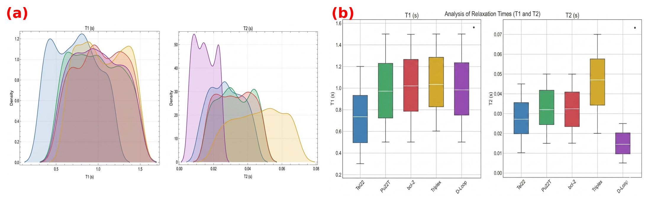
Rare DNA intermediate structures are essential for biological processes in living organisms, including protein folding and unfolding, gene transcription, and replication. Studies have been conducted on DNA intermediate structures using different nuclear magnetic resonance (NMR) approaches, but NMR-based statistical analysis of DNA is less researched. Therefore, the current study presents a statistical analysis of NMR longitudinal (T1) and transverse (T2) relaxation times for rare DNA intermediate structures. Published experimental T1 and T2 datasets for five rare DNA intermediate structures were simulated using an R script to obtain 500 data points for each DNA structure; these were used to perform non-parametric statistical analysis using Wolfram Mathematica software. Results of the analysis show that the T1 and T2 datasets for each of the five rare DNA structures have distinct features. This highlights the possibility of utilising DNA T1 and T2 NMR parameters to isolate DNA structures responsible for regulating gene transcription, replication, and repair.
References
[1] J. Sun, Y. Wang, Y. Huang & Z. Chen, ``G-quadruplex unwinding molecular mechanisms by helicases and their applications'', International Journal of Molecular Sciences 27 (2026) 1629. https://doi.org/10.3390/ijms27041629.
[2] E. Soorya & S. S. Pingale, ``DFT analysis of stacking interactions and cyclic H-bond cooperativity in G-quadruplexes'', Journal of Computational Chemistry 46 (2025) e70290. https://doi.org/10.1002/jcc.70290.
[3] A. L. Soenarjo, Z. Lan, I. V. Sazanovich, Y. S. Chan, M. Ringholm, A. Jha & D. R. Klug, ``The transition from unfolded to folded G-quadruplex DNA analyzed and interpreted by two-dimensional infrared spectroscopy'', Journal of the American Chemical Society 145 (2023) 19622. https://doi.org/10.1021/jacs.3c04044.
[4] J. Y. Gong, C. J. Wen, M. L. Tang, R. F. Duan, J. N. Chen, J. Y. Zhang, K. W. Zheng, Y. D. He, Y. H. Hao, Q. Yu, S. P. Ren & Z. Tan, ``G-quadruplex structural variations in human genome associated with single-nucleotide variations and their impact on gene activity'', Proceedings of the National Academy of Sciences of the United States of America 118 (2021) e2013230118. https://doi.org/10.1073/pnas.2013230118.
[5] S. Rhee, Z. J. Han, K. Liu, H. T. Miles & D. R. Davies, ``Structure of a triple helical DNA with a triplex-duplex junction'', Biochemistry 38 (1999) 16810. https://doi.org/10.1021/bi991811m.
[6] M. D. Frank-Kamenetskii & S. M. Mirkin, ``Triplex DNA structures'', Annual Review of Biochemistry 64 (1995) 65. https://doi.org/10.1146/annurev.bi.64.070195.000433.
[7] I. Kovalchuk & O. Kovalchuk (Eds.), Genome Stability: From Virus to Human Application, 2nd edition, Academic Press, Amsterdam, Netherlands, 2021, pp. 117--131. https://doi.org/10.1016/C2020-0-00641-9.
[8] J. Robinson, F. Raguseo, S. P. Nuccio, D. Liano & M. Di Antonio, ``DNA G-quadruplex structures: more than simple roadblocks to transcription?'', Nucleic Acids Research 49 (2021) 8419. https://doi.org/10.1093/nar/gkab609.
[9] L. Joudeh, R. E. Appleby, J. D. Maman & L. Pellegrini, ``Structural mechanism of strand exchange by the RAD51 filament'', eLife 14 (2025) RP107114. https://doi.org/10.7554/eLife.107114.
[10] B. H. Annex & R. S. Williams, ``Mitochondrial DNA structure and expression in specialized subtypes of mammalian striated muscle'', Molecular and Cellular Biology 10 (1990) 5671. https://doi.org/10.1128/mcb.10.11.5671-5678.1990.
[11] J. N. Doda, C. T. Wright & D. A. Clayton, ``Elongation of displacement-loop strands in human and mouse mitochondrial DNA is arrested near specific template sequences'', Proceedings of the National Academy of Sciences of the United States of America 78 (1981) 6116. https://doi.org/10.1073/pnas.78.10.6116.
[12] J. M. Yang, J. Kim, Y. Lee, W. Lee & J. C. Park, ``NMR structure and biophysical characterization of thermophilic single-stranded DNA binding protein from Sulfolobus solfataricus'', International Journal of Molecular Sciences 23 (2022) 3099. https://doi.org/10.3390/ijms23063099.
[13] C. A. Brosey, S. E. Soss, S. Brooks, C. Yan, I. Ivanov, K. Dorai & W. J. Chazin, ``Functional dynamics in replication protein A DNA binding and protein recruitment domains'', Structure 23 (2015) 1028. https://doi.org/10.1016/j.str.2015.04.008.
[14] C. Brown, K. Campos-Leon, M. Strickland, C. Williams, V. S. S. Fairweather, R. L. Brady, M. P. Crump & K. Gaston, ``Protein flexibility directs DNA recognition by the papillomavirus E2 proteins'', Nucleic Acids Research 39 (2011) 2969. https://doi.org/10.1093/nar/gkq1217.
[15] L. E. Thompson & D. Rovnyak, ``Accessible NMR experiments studying the hydrodynamics of $^{15}$N-enriched ubiquitin at low fields'', Biochemistry and Molecular Biology Education 35 (2007) 49. https://doi.org/10.1002/bmb.23.
[16] C. Castagn'e, E. C. Murphy, A. M. Gronenborn & M. Delepierre, ``$^{31}$P NMR analysis of the DNA conformation induced by protein binding: SRY/DNA complexes'', European Journal of Biochemistry 267 (2000) 1223. https://doi.org/10.1046/j.1432-1327.2000.01124.x.
[17] R. Sahu, A. Kumar, R. Mishra, C. Majee & R. Kumar, ``Recent development in nuclear magnetic resonance as a technique for quantitative method validation'', Indian Journal of Pharmaceutical Sciences 83 (2021) 1094. https://doi.org/10.36468/pharmaceutical-sciences.865.
[18] O. B. Awojoyogbe, M. O. Dada, O. P. Faromika & O. E. Dada, ``Mathematical concept of the Bloch flow equations for general magnetic resonance imaging: A review'', Concepts in Magnetic Resonance Part A 38A (2011) 85. https://doi.org/10.1002/cmr.a.20210.
[19] E. Bairaktari, K. Katopodis, K. C. Siamopoulos & O. Tsolas, ``Paraquat-induced renal injury studied by $^1$H nuclear magnetic resonance spectroscopy of urine'', Clinical Chemistry 44 (1998) 1256. https://doi.org/10.1093/clinchem/44.6.1256.
[20] R. W. Darbeau, ``Nuclear magnetic resonance (NMR) spectroscopy: A review and a look at its use as a probative tool in deamination chemistry'', Applied Spectroscopy Reviews 41 (2006) 401. https://doi.org/10.1080/05704920600726175.
[21] G. Patel & D. Chudasama, ``Nuclear magnetic resonance spectroscopy NMR'', Journal of Nuclear Engineering & Technology 11 (2021) 30. https://www.researchgate.net/publication/357735866_Nuclear_Magnetic_Resonance_Spectroscopy_NMR.
[22] D. Marion, ``An introduction to biological NMR spectroscopy'', Molecular & Cellular Proteomics 12 (2013) 3006. https://doi.org/10.1074/mcp.O113.030239.
[23] P. J. Sapienza & A. L. Lee, ``Using NMR to study fast dynamics in proteins: methods and applications'', Current Opinion in Pharmacology 10 (2010) 723. https://doi.org/10.1016/j.coph.2010.09.006.
[24] A. Ambrus & D. Yang, ``Diffusion-ordered nuclear magnetic resonance spectroscopy for analysis of DNA secondary structural elements'', Analytical Biochemistry 367 (2007) 56. https://doi.org/10.1016/j.ab.2007.04.025.
[25] H. Abou Assi, M. Garav'is, C. Gonz'alez & M. J. Damha, ``i-Motif DNA: structural features and significance to cell biology'', Nucleic Acids Research 46 (2018) 8038. https://doi.org/10.1093/nar/gky735.
[26] Y. Huang, Development of antisense alpha-helical peptide nucleic acids, Ph.D. dissertation, Department of Chemistry, Case Western Reserve University, Cleveland, Ohio, 2002. https://www.proquest.com/openview/837b212f801191c2059c70bc780e04e8/1?pq-origsite=gscholar&cbl=18750&diss=y.
[27] D. M. J. Lilley & D. G. Norman, ``The Holliday junction is finally seen with crystal clarity'', Nature Structural Biology 6 (1999) 897. https://doi.org/10.1038/13255.
[28] A. Bansal, S. Kaushik & S. Kukreti, ``Non-canonical DNA structures: diversity and disease association'', Frontiers in Genetics 13 (2022) 959258. https://doi.org/10.3389/fgene.2022.959258.
[29] F. Y. Teng, Z. Z. Jiang, M. Guo, X. Z. Tan, F. Chen, X. G. Xi & Y. Xu, ``G-quadruplex DNA: a novel target for drug design'', Cellular and Molecular Life Sciences 78 (2021) 6557. https://doi.org/10.1007/s00018-021-03921-8.
Published
How to Cite
Issue
Section
Copyright (c) 2027 John A. Lazarus, Michael O. Dada, Awojoyogbe O. Bamidele, Simon O. Abolarinwa (Author)

This work is licensed under a Creative Commons Attribution 4.0 International License.