Deep convolutional neural network (DCNN)-based model for pneumonia detection using chest x-ray images

Authors

  • S. I. Ele Department of Computer Science, University of Calabar, P. M. B 1115, Etagbor, Cross River State, Nigeria
  • U. R. Alo Computer Science Department, Alex Ekwueme Federal University, Ndufu-Alike, P.M.B 1010, Abakaliki, Ebonyi State, Nigeria https://orcid.org/0000-0001-5196-764X
  • H. F. Nweke Computer Science Department, David Umahi Federal University of Health Science, P.M.B 211, Uburu, Ebonyi State, Nigeria https://orcid.org/0000-0001-5196-764X
  • A. H. Okemiri Computer Science Department, Alex Ekwueme Federal University, Ndufu-Alike, P.M.B 1010, Abakaliki, Ebonyi State, Nigeria
  • E. O. Uche-Nwachi Computer Science Department, Alex Ekwueme Federal University, Ndufu-Alike, P.M.B 1010, Abakaliki, Ebonyi State, Nigeria

Keywords:

Machine Learning , Convolutional Neural Network (CNN), Artificial Intelligence, Pre-trained Models, Pneumonia Detection

Abstract

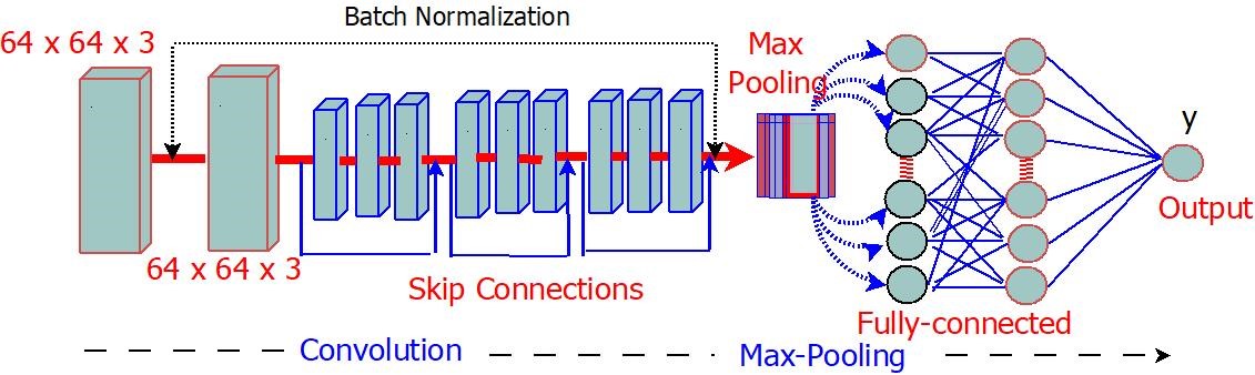
In recent years, the integration of machine learning techniques within the medical field has shown promising results in aiding healthcare pro[1]fessionals in accurate diagnosis and treatment planning. This study focuses on developing and implementing a machine learning model tailored specifically for medical diagnosis, leveraging advancements in computer vision and deep learning algorithms. This research aims to design an efficient and accurate model capable of classifying medical images into distinct categories, enabling automated diagnosis and identification of various ailments and conditions. This study uses a dataset comprising 5,863 Chest X-ray images (JPEG) and 2 categories (Pneumonia/Normal) (anterior-posterior) selected from retrospective cohorts of pediatric patients of one to five years old from Guangzhou Women and Children’s Medical Center, Guangzhou, obtained from Kaggle data repositories. Data Preprocessing was conducted to enhance image quality and extract relevant features, followed by implementing a deep convolutional neural networks (DCNNs) model using TensorFlow’s Keras. Using pre-trained models such as Resnet, transfer learning techniques were employed to learn efficient features from large-scale datasets and optimize the model’s performance with the limited medical data available. The results from the experimental analysis showed that after 9 epochs, the training and validation accuracies had steadily increased, achieving 95% and 75%, respectively. Overall, the model achieved 99.9% training accuracy across multiple epochs and an average validation accuracy of 75%. The model’s performance and scalability highlight its potential for integration into clinical workflows. This could revolutionize healthcare by augmenting the diagnostic process and improving patient outcomes.

Dimensions

[1] Pneumonia in children, by World Health Organization. (2022, Novem ber 11). [Online]. https://www.who.int/news-room/fact-sheets/detail/pneumonia.

[2] Pneumonia, by Centers for disease control and prevention. (2022). [Online]. https://www.cdc.gov/pneumonia/index.html.

[3] T. Vos, S. S. Lim, C. Abbafati, K. M. Abbas, M. Abbasi, M. Abbasi fard, M. Abbasi-Kangevari, H. Abbastabar, F. Abd-Allah, A. Abdelalim & M. Abdollahi, “Global burden of 369 diseases and injuries in 204 countries and territories, 1990–2019: a systematic analysis for the Global Burden of Disease Study 2019”, The Lancet 396 (2023) 1222. https://www.thelancet.com/journals/lancet/article/piis0140-6736(20)30925-9/fulltext.

[4] A. H. Mokdad, K. Ballestros, M. Echko, S. Glenn, H. E. Olsen, E. Mullany, A. Lee, A. R. Khan, A. Ahmadi, A. J. Ferrari & A. Kasaeian, “The state of US health, 1990-2016: Burden of diseases, injuries, and risk factors among US states”, JAMA-Journal of the American Medical Association 319 (2018) 1472. https://doi.org/10.1001/jama.2018.0158.

[5] T. O’Driscoll, “The economic burden of pneumonia in the United States: An analysis of national databases”, European Respiratory Journal 52 (2018) 1801582. https://doi.org/10.1183/13993003.01582-2018.

[6] M. El-Ghandour & M. I. Obayya, “Pneumonia detection in chest x-ray images using an optimized ensemble with XGBoost classifier”, Multimedia Tools and Applications (2024) 1. https://doi.org/10.1007/s11042-024-18975-6.

[7] V. Chouhan, “A novel transfer learning based approach for pneumonia detection in chest X-ray images”, Applied Sciences 10 (2020) 559. https://www.mdpi.com/2076-3417/10/2/559.

[8] M. Togac¸ar, B. Ergen, Z. C ? omert & F. ¨ Ozyurt, “A deep feature learning model for pneumonia detection applying a combination of mRMR feature selection and machine learning models”, Irbm 41 (2020) 222. https://doi.org/10.1016/j.irbm.2019.10.006.

[9] O. Bwanga, “Barriers to continuing professional development (CPD) in radiography: a review of literature from Africa”, Health Professions Ed ucation 6 (2020) 480. https://doi.org/10.1016/j.hpe.2020.09.002.

[10] Z. Gilani, Y. D. Kwong, O. S. Levine, M. Deloria-Knoll, J. A. G. Scott, K. L. O’Brien & D. R. Feikin, “A literature review and survey of childhood pneumonia etiology studies: 2000–2010”, Clinical Infectious Diseases 54 (2012) S108. https://doi.org/10.1093/cid/cir1053.

[11] R. E. Black, “Global, regional, and national causes of child mortality in 2008: a systematic analysis”, Lancet 375 (2010) 87. https://doi.org/10.1016/S0140-6736(10)60549-1.

[12] Integrated Management of Childhood Illness: Chart Booklet, by World Health Organization. (2014, March 4). [Online]. https://www.who.int/publications/m/item/integrated-management-of-childhood-illness---chart-booklet-(march-2014).

[13] Global Burden of Disease Study 2019, by Institute for Health Metrics and Evaluation. (2019). [Online]. http://ghdx.healthdata.org/gbd-2019.

[14] Z. Feng, Q. Yu, S.Yao, L. Luo, W. Zhou, X. Mao & W. Wang, “Early prediction of disease progression in COVID-19 pneumonia patients with chest CT and clinical characteristics”, Nature communications 11 (2020) 4968. https://doi.org/10.1038/s41467-020-18786-x.

[15] P. Rajpurkar, J. Irvin, K. Zhu, B. Yang, H. Mehta, T. Duan, D. Ding, A. Bagul, C. Langlotz & K. Shpanskaya, “Chexnet: radiologist-level pneumonia detection on chest x-rays with deep learning”, arXiv preprint arXiv:1711.05225. https://stanfordmlgroup.github.io/projects/chexnet/.

[16] T. B. Chandra & K. Verma, Pneumonia detection on chest x-ray using machine learning paradigm, Proceedings of 3rd International Conference on Computer Vision and Image Processing: CVIP 2018, Singapore, 2020. https://doi.org/10.1007/978-981-32-9088-4 3.

[17] M. Togac¸ar, B. Ergen, Z. C ? omert & F. ¨ Ozyurt, “A deep feature learning model for pneumonia detection applying a combination of mRMR feature selection and machine learning model”, IRBM 41 (2022) 222. https://doi.org/10.1016/j.irbm.2019.10.006.

[18] D.S. Kermany, M. Goldbaum, W. Cai, C. C. Valentim, H. Liang, S. L. Baxter & K. Zhang, “Identifying medical diagnoses and treatable diseases by image-based deep learning”, Cell 172z (2018) 1131. https://doi.org/10.1016/j.cell.2018.02.010.

The structure of the DCNN pneumonia detector system.

Published

2025-05-01

How to Cite

Deep convolutional neural network (DCNN)-based model for pneumonia detection using chest x-ray images. (2025). Journal of the Nigerian Society of Physical Sciences, 7(2), 2443. https://doi.org/10.46481/jnsps.2025.2443

Issue

Section

Computer Science

How to Cite

Deep convolutional neural network (DCNN)-based model for pneumonia detection using chest x-ray images. (2025). Journal of the Nigerian Society of Physical Sciences, 7(2), 2443. https://doi.org/10.46481/jnsps.2025.2443